
MRI technology has come a long way since its inception in the early 1970s. Magnetic resonance imaging, or MRI, is a medical diagnostic procedure that uses a strong magnetic field and radiofrequency pulses to produce images of the body. The first MRI scanner was built by Dr. Raymond Damadian in 1971, and it took nearly a decade for the technology to be refined enough for widespread use. Despite these early challenges, it has become an invaluable tool in modern medicine, providing physicians with detailed images of internal organs and tissues without the need for surgery or radiation, with companies such as Excedr leading the way with the technology.
How MRI Works
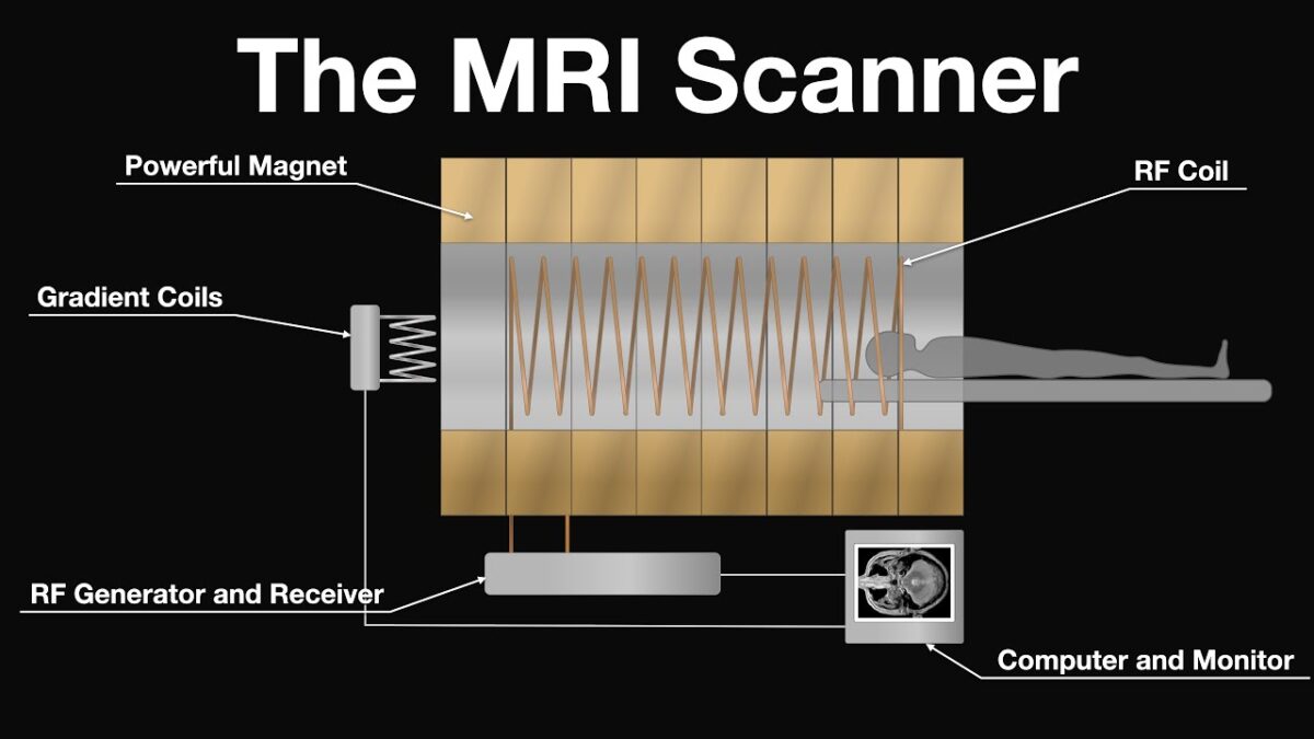
It works by detecting the natural energy emitted from hydrogen atoms in different body tissues. These hydrogen atoms have a nucleus with a single positive charge and a single unpaired electron spinning around that charge. When placed into an external magnetic field, the nucleus of these hydrogen atoms will align itself according to that field’s polarity, just as a compass needle is drawn to the Earth’s North Pole. When the body is placed into a strong magnetic field, these hydrogen nuclei are polarized and spin at their resonance frequency.
The nuclei of these atoms will also be slightly affected by radio waves transmitted through the body. When these radio waves are turned off and the nuclei return to their normal state, they give off a radio frequency signal detected by an array of receiver coils and transmitted into real-time images. This process will be repeated for different angles of the body, which results in three-dimensional images of the organs and tissues inside the patient’s body.
History of MRI Technology
The first image from an MRI scanner was produced in 1977, just a short time after the first commercial scanners were made available to hospitals and clinics. Then, in 1982, Damadian published results showing that tumors could be detected earlier using the technology. Still, it would take several more years before larger studies confirmed his findings. After that, MRI technology continued to make strides, eventually culminating in the first full-body 3D scan in 1998.
Commercial Market Implementation
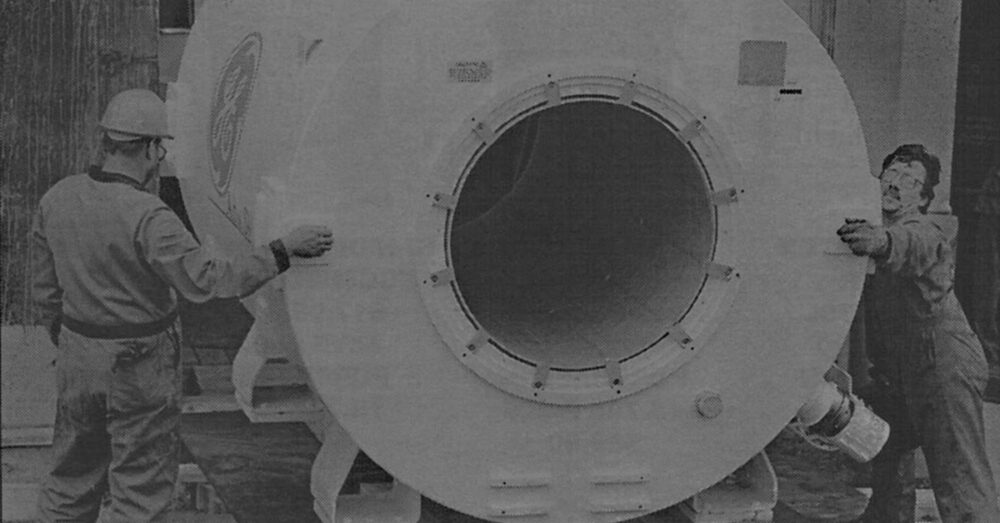
The prototype of an MRI machine was built at New York University Medical Center by Dr. Damadian and his colleagues in 1971. Still, it would take until 1977 before the first commercial MRI scanner would be available to physicians. Many technical difficulties had to be overcome before this could happen, but the introduction of higher-strength permanent magnets made it possible by enhancing image quality and resolution. It was still several years before other companies started producing their versions of the technology at an affordable price for hospitals and clinics worldwide. Still, today several companies have a significant market share in the MRI scanning industry, including GE Healthcare and Hitachi.
What MRI Can Be Used To Diagnose
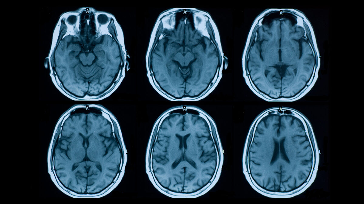
MRI can be used to examine almost any part of the body, including organs, soft tissues, and bones. The most common types of scans that are performed today include:
- T1-weighted images, which provide information about the structure of different tissues.
- T2-weighted images, which give physicians an idea about how healthy those tissues are.
- Diffusion imaging, which can highlight specific structures with impaired blood flow.
- Functional imaging, which can show brain activity by tracking blood flow and oxygen levels in the body.
- 3D-imaging, which allows physicians to examine specific structures in three dimensions.
What MRI Cannot Be Used To Diagnose
MRI has become an invaluable tool for medical professionals worldwide, but there are still some conditions it cannot be used to detect. For instance, MRI scans do not work well for diagnosing neurological diseases because the hydrogen atoms in water molecules scatter the radio waves instead of aligning themselves underneath the magnetic field. This makes imaging deep into parts of the brain or spine very difficult, although some scanning sequences can pick up on certain abnormalities. In addition, MRI is not a good way to detect many types of infections, such as the early stages of tuberculosis, until it has reached a more advanced stage.
How an MRI Scan Is Performed
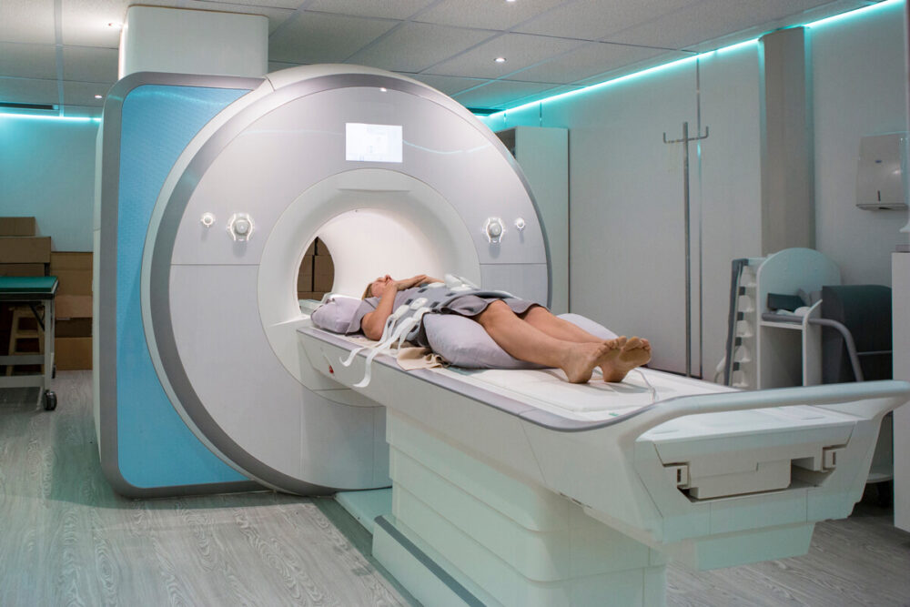
The typical MRI procedure will start with the patient being asked to remove all metal objects from their person. This is because the powerful magnets used in MRI scanners can cause these objects to injure the patient. Once the patient is inside the scanning machine, they will be asked to lie still on their back or stomach as different images are taken. Depending on the part of the body that is being scanned, the procedure can take anywhere from a few minutes to half an hour.
MRI Scanners
There are two main types of MRI scanners: open and closed. Open scanners have a large opening in the front or back that allows patients to be placed inside without having to go through a tunnel. This type of scanner is often used for scanning children or patients who are claustrophobic. Closed MRI scanners have a much smaller opening and patients must be placed inside a tight tunnel in order to be scanned. This type of scanner is more common, as it provides higher-quality images.
Controversy in Medicine
Since it was first introduced into medical practices all over the world, there have been disputes about whether or not MRI scans are safe for patients. In particular, there has been some concern about whether or not MRI technology can interfere with the devices that are used to implant pacemakers and inserted metallic prosthetics. However, studies have shown that patients who receive a standard MRI scan will not be affected by magnetic forces as long as they do not have any electronic implants.
Another controversy that has arisen around MRI technology is the use of gadolinium-based contrast agents. These are used to make body tissues more visible on an MRI scan, but there have been some concerns that they could be harmful to patients’ health. So far, there is no concrete evidence that these agents cause any long-term health problems, but further research is still needed.
Closing Thoughts on the History of MRI Technology
Magnetic resonance imaging has become one of the most important tools for medical professionals worldwide, allowing them to diagnose and treat problems that would otherwise be undetectable. Although it wasn’t always available for use in hospitals and clinics, the first commercial scanner was introduced in 1977, and physicians have been using MRI scans ever since. MRI technology has been revolutionary for medicine, but it will likely undergo some changes over the years as the demand for better technology increases.


